Nervous System Primer
Neuron anatomy
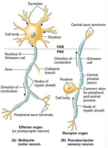
Cell body is where the nucleus resides. Many cell bodies together is a ganglion in the PNS and a nucleus in the CNS.
Dendrites: Receive signal
Axon: Send signal

Nerves
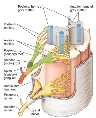
Nerves reside in the horn roots 😈 of gray matter. Rootlets emerge from horns.
Sensory neuron in dorsal horns. On-ramps.
Motor nuclei reside ventral horns. Off-ramps.
Rootlets emerge from horns and form dorsal/ventral roots. These combine to form spinal nerves which branch. Spinal nerves branch into rami, the dorsal ramus and ventral ramus. Because it is both dorsal and ventral, it has both motor and sensory info.
Dorsal ramus: Intrinsic muscles of back and skin of back
Ventral ramus: Everything else
The dorsal root ganglion is a good way of finding dorsal direction.

Spinal nerves
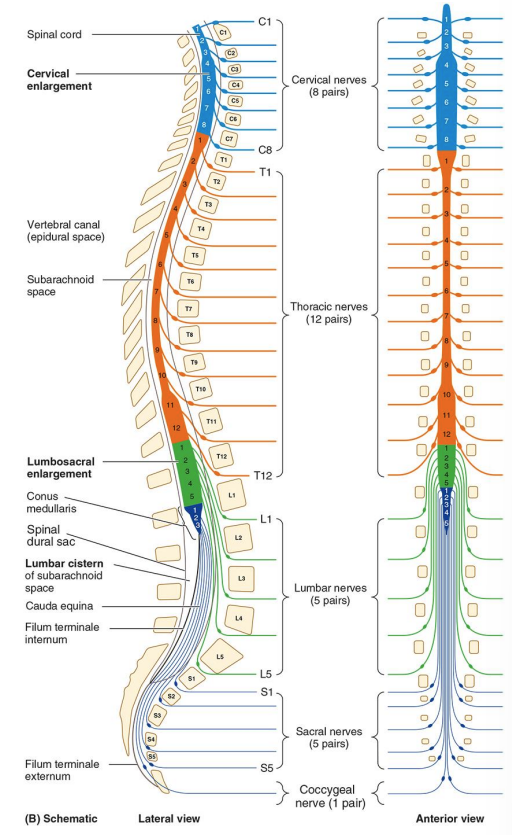
Named by vertebral region and the number in region.
Spinal cord ends at L1/L2. The conus medularis is the official end to the spinal cord. The filum terminale emerges from the tip of the conus medularis and continues as the cauda equina.


Dermatomes
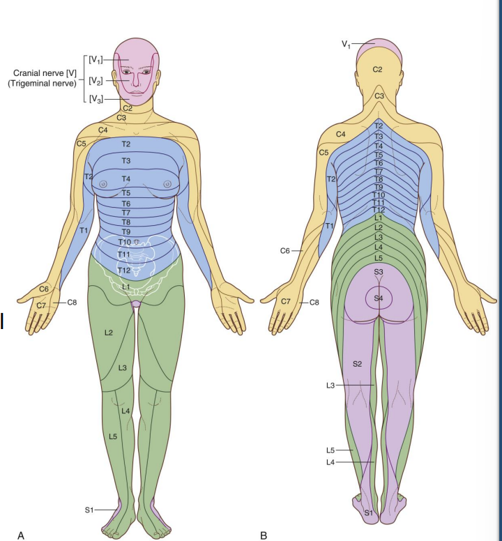
Provide sensory innervation to the skin. Mostly ventral rami. These will follow the naming vertebrae of the spine. A rash following a particular dermatome may indicate pathology of the corresponding spinal nerve (e.g. shingles).
There are no dermatomes for the face as sensory information for the head and neck is picked up by cranial nerves. The exception to this is cranial nerve X (vagus nerve) which goes everywhere (heart, lungs, GI, etc.)

Autonomic Nervous System
Sympathetic: Thoracolumbar (T1-L2)
Parasympathetic: Cranial nerves (III, VII, IX, X) & S2-4 spinal segments
- Preganglionic cell
- Autonomic ganglion
- Post ganglionic cell
- Post ganglionic neuron
- Target

Sympathetic
Is responsible for big, stressful responses. Activation will increase blood pressure via innervation of blood vessels.
Sensory signals can only be innervated in lateral horn of T1-L2. However, to get innervation elsewhere in the body, signals travel from the ventral (motor) root of the spinal nerve. This central spinal nerve connection is called the white ramus communicans.
Paravertebral ganglia (sympathetic trunk): Sympathetic ganglion running vertically along the spinal cord, connecting at each segment
Prevertebral ganglia: SNS ganglia located away from sympathetic chain ganglia at the anterior midline of the aorta
Pathway
From brain → body
- Lateral horn (T1-L2)
- Ventral root (joins with Dorsal root) to form spinal nerve
- White ramus communicans (myelinated)
- Sympathetic chain options
- Synapse at same level
- Paravertebral ganglia: Ascend/synapse OR descend/synapse
- Gray ramus communicans (unmyelinated)
- Connect back to spinal nerve
- Ventral or dorsal ramus (depending on body location target)
- Leave chain without synapsing (splanchnic nerve) which will go on to synapse at prevertebral ganglia
Gray (off ramps) are all along the sympathetic trunk. White (on ramps) only exist at T1-L2.
Gray all the way, white not quite
For more on white and gray matter, see here: Neuroscience Concepts > White and gray matter
Sympathetic splanchnic nerves
- Greater (T5-T9)
- Lesser (T10-T11)
- Least (T12)
- Lumbar (L1-L2)
- Exit in lumbar region
- Sacral (L1-L2)
- Exit in sacral region
These will send signals to very specific innervation sites. The lesser splanchnic nerve also supplies the midgut.
Referred pain
Visceral (organ) pain neurons often wire together with sympathetic fibers. The neuron will send a signal along these routes rather than having its own pathway. Therefore, pain will be localized to the region (diffuse) as it activates the entire dermatome. Somatic pain (e.g. pinching the skin), is more specific.
Parasympathetic
Preganglionic fibers in the PSNS are usually pretty long and synapse directly with the target organ. The vagus nerve (cranial nerve X) is an example of this and will synapse with short post-ganglionic neurons at the end organs of the thorax and abdomen.
The peripheral parasympathetic ganglion primarily targets the head.
Intermural ganglion (usually rather short): In the walls of the target organ connect connect pre and postganglionic cells
Preganglionic parasympathetic nerves
Cranio
Nuclei of cranial nerves III (oculomotor), VII (facial), IX (glossopharyngeal), and X (vagus)
Brain Stem → Vagus Nerve (CN X)→ Organs in Thorax and Abdomen
Sacral
Lateral horns of S2-4 (travel along the pelvic spanchnics)
Sacral → Pelvic Nerves → Distal Colon, Bladder, Reproductive Organs