Circulatory System Primer
For circulatory system development, see here: Mesoderm - Cardiovascular
Cardiovascular system
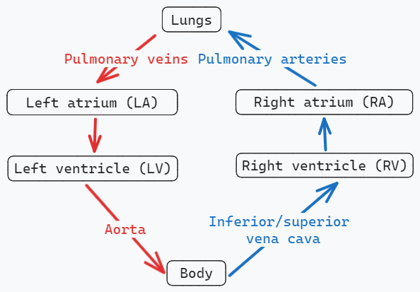
Blood vessels
Arteries
- Thick smooth muscle
- Usually high oxygen
- Arterioles deliver blood to capillaries
Types of arteries
- Large elastic (conducting) arteries: Elastic layers which control pressure changes (e.g. aorta + primary branches, pulmonary trunk + branches)
- Medium muscular arteries: Circular smooth muscle allows for vasoconstriction. These regulate blood flow. These make up the majority of named arteries
- Small arteries and arterioles: Generally not named
Capillaries
- Form capillary beds
- Exchange oxygen, nutrients, fluids, waste products, etc.. with the extracellular fluid
Veins
- Venules drain capillaries
- Low oxygen
- Flexible walls
Types of veins
- Large veins: The vena cava
- Medium (superficial) veins: Drain venus plexuses (e.g. cephalic and basilic veins)
- Small veins: Small veins combine to form venous plexuses
- Venules: drain into capillary beds
Telling arteries from veins: Arteries hold their shape, whereas veins flatten out and have wrinkly thin walls.
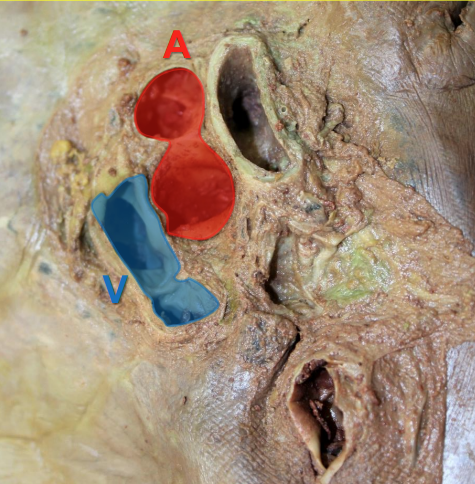
Large arteries need elasticity in order to conform to pressure from heart. Medium muscular arteries allow for vasoconstriction/dilation to regular blood flow.
Anastomoses
Connections between artery branches. Provide collateral circulation (alternate pathways) to regions/organs which prevents ischemia as other pathways will compensate. In some cases, may get retrograde flow. Common around joints.
Vein concepts
From small veins you have drainage, or movement back to the heart. Most drainage veins have names.
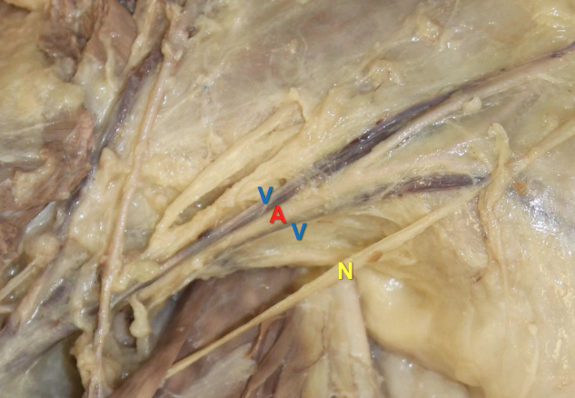
In many cases, medium veins will diverge into two or more branches which accompany a deep artery (usually on either side). This is referred to as venae comitantes. The VC is wrapped in a fascia sheathe. Expansion of the arteries stretch and flatten the veins, acting as an arteriovenous pump which drives the blood back to the heart.
Plexus: Intricate network
Telling arteries from veins and nerves
Arteries and veins share a fascial sheath. Nerves will originate from elsewhere
Lymphatic system
Carries lymph from interstitial spaces to venous system, filtering the blood. Is a key part of the innate immune response.
Lymphatic capillaries: Points where fluid/particulates absorb
Lymphatic vessels: Carries lymph from capillaries to venous system
Lymph nodes: Masses of lymph. Lymph is filtered through these on its way to venous system
Lymphocytes: Immune (B and T) cells
Lymphoid organs: Produces, matures, or activates lymphocytes. (Thymus, red bone marrow, spleen, tonsils, lymph nodules)
Leaky capillaries
The pressure in capillary beds is higher than in interstitium, which drives fluid into the latter. 3L per day.
Lymphatic vessels
A single later of overlapping epithelial cells makes up lymphatic vessels. This allows for easy transference of debris/bacteria/etc. to lymphatic system. Drainage tends to flow in the direction of venous drainage.
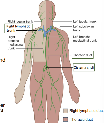
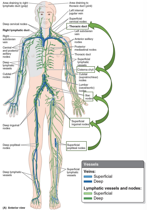
Right lymphatic duct drains the right upper limb / right side of head, neck, and thorax at confluence of right internal jugular and right subclavian veins.
Thoracic duct drains into the rest of the body at the confluence of let internal jugular and left subclavian vein.
Cisternal chyli drains from the lower limbs, pelvis, and abdomen into thoracic duct
"Chyle" comes from the high fat lymph in the intestines
Basically, if its not draining into the top part of the body, it is for sure draining into the thoracic duct.
Lymph nodes
Named for region, organ, or blood vessel.
Superficial (e.g. cutaneous) nodes often names for venous drainage. Lymphatic vessels deep in the gut (e.g. abdominal organs) are often named for the arteries there as they flow in the liver direction.
You should be able to read the name and determine direction of movement.
Sentinel nodes
The first lymph node where cancer cells may spread into a tumor from an organ. Lymph is highly anastomotic (multiple directions the lymph can drain). When removing tumors, the lymph node is also sometimes removed from regions known to drain that organ. This can determine the presence or extent of metastasis depending on goals of surgery.
For exam details, see here: Lymph Node Exam