Cardiopulmonary Exam
Cardio Exam: Step-by-step
- Adjust table to a 45 deg angle (opening the foot rest) and stand to patient's right side
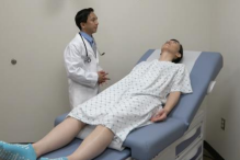
- Drape patient so as to access the left side of their chest
- Inspect precordium: Palpate 2nd, 3rd, and 4th intercostal spaces (right and left side of sternum)
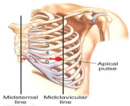
- Palpate PMI (Point of Maximal Impulse): 5th ICS/midclavicular
- The patient may have to raise their left breast to access this
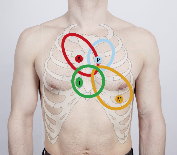
- Auscultate heart with steth (All Physicians Take Money). Use Diaphragm to hear S1 ("lub") and S2 ("dub") sounds.
- Aortic: 2nd ICS/right sternal border
- Pulmonic: 2nd ICS/left sternal border
- Tricuspid: 5th ICS/midclavicular line
- Mitral: 5th ICS/midclavicular line
- Repeat with bell
- Tricuspid: 5th ICS/midclavicular line
- Mitral: 5th ICS/midclavicular line
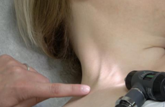
- Palpate carotid arteries: Bilaterally, one at a time (lol)
- Auscultate carotid arteries (diaphragm)
- Have patient to hold breath when listening
- Listen at medial border of SCM (diaphragm)
- Ask patient to hold breath
- Check jugular venous pressure
- Identify
- Locate top of jugular pulsations (meniscus)
- Place vertical ruler at sternum
- Place straight edge at top of jugular, intersecting with ruler perpendicularly
- Calculate JVP
- Measure vertical height on ruler and add 5cm
- 6-8 cm H2O is normal
- Check bilateral radial pulses in wrists
- Check bilateral dorsalis pedis (top of foot) pulses
- Check for swelling. Firm pressure with thumbs looking for indentation in 3 areas (bilaterally):
- Dorsum of foot
- Lower tibia
- Upper tibia
Pulm Exam: Step-by-step
- Ask patient to expose back
- Inspect shape of chest rise
- Observe breathing rate, effort, and muscle use
- Respiratory expansion (normal rate = 12-16 BPM)
- Place hands on mid back, around lower rib 10
- Ask patient to take deep breath
- Assess chest excursion, checking for symmetry
- Percuss posterior lung fields (horizontal and then vertical, to compare sides)
- Place hand on back, tapping firmly on middle finger
- 5 posterior and 2 lateral levels. Bilaterally and symmetrically, avoiding scapula
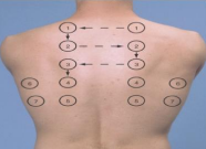
- Percuss anterior lung fields: 2 levels
- 1 above clavicle and 1 below clavicle
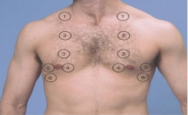
- 1 above clavicle and 1 below clavicle
- Auscultate lungs
- Ask patient to take deep breaths though open mouth
- 5 posterior and 2 lateral levels (same as above)
- 1 above clavicle and 1 below clavicle (same as above)
Note on steth
Diaphragm: Good for all frequencies
Bell: Good for medium and low frequencies