HEENT And Neck Exam
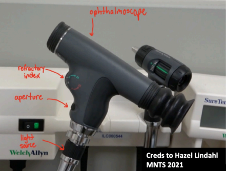
Ophthalmoscope
Has three dials:
- Light source - Controls the light intensity. Medium is usually fine.
- Aperture - Use the medium circle (green indicator)
- Refractive index - Adjusts for corrective factor. Leave setting at 0 for no glasses.
Otoscope
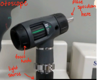
Has two dials:
- Light source (same as ophthalmoscope)
- Focus knob (green line indicates neutral position)
Surface Anatomy
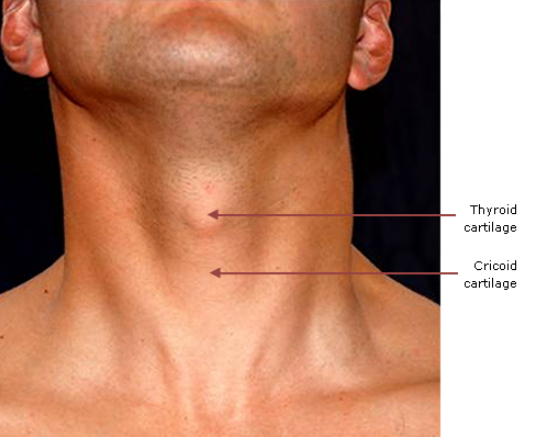
Place finger on middle of patient's chin and move down.
1st structure = thyroid cartilage
2nd structure = cricoid cartilage
3rd structure = isthmus of the thyroid (just inferior to cricoid)
Palpating the thyroid
- First, ask patient to swallow and feel the movement.
- Next, place both hands on isthmus from behind
- Slightly rock the cartilage back and fourth, feeling each lobe
- Use one hand to fix the thyroid in place and walk fingers down lobe. Once for left and right
Head exam
- Inspect head
- Inspect scalp and hair

Eye exam
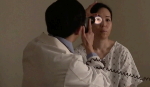
- Pull down lower lids and ask patient to look up. Note any abnormal coloration of conjunctivae, sclera, and pupils.
- Dim lights and prep ophthalmoscope
- Ask patient to focus on fixed point in distance
- Stabilize your hand on forehead and position yourself arms length away
- Approach nearer to patient's eye from slight lower angle until eye cup touches patient's brow
- Identify blood vessels and optic disc. Note spots, damaged blood vessels, and abnormal vessel formation
Ears
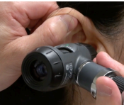
- Inspect ears, noting lumps or fluid
- Place speculum tip on otoscope
- Straighten patient's ear by gently pulling up the pinna
- Insert the tip and inspect patient's tympanic membrane. Note if membrane is not pearly gray, translucent, and shiny
- Repeat for other ear

Nose
- Place speculum top on otoscope and ask patient to tilt back head
- Insert otoscope into each nostril (naris) and inspect mucosa, septum, turbinate for inflammation
- Palpate sinuses and ask if the patient feels any pain

Mouth
- Use light source and tongue blade to inspect gums, teeth, tongue, and oral mucosa
- Ask patient to lift tongue and inspect subglossal area
- Ask patient to stick out tongue say "Ah" while pressing down on tongue
Lymph nodes
- Inspect lymph nodes
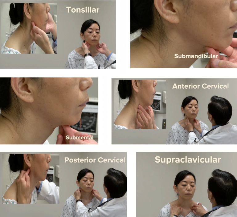
- Palpate six regions: tonsillar, submandibular, submental, anterior cervical, posterior cervical, supraclavicular. Note swelling or tenderness.
