Ultrasound
In which a machine yells into your body (quietly) and makes a picture based on many sound waves bounce back. Soft tissues will have a weaker echo and thus the machine ear (transducer) can discriminate anatomical features.
It's worth reviewing the spatial anatomical terms before conducting an ultrasound: Anatomy Nomenclature
Artifacts
An image from faulty wave interpretation.
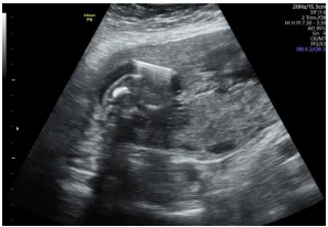
Reverberation
Waves bounce between two hard surfaces. Requires an angle change, more gel, or patient repositioning. Seen below as the white line with reverberation underneath.
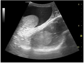
Mirror image
Double the image, double the fun. The real one is usually closer to the transducer. Requires a change in probe angle.
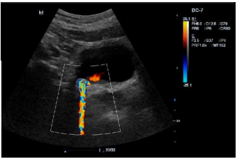
Twinkle
Waves will sometimes reflect of a calcified object and cause it to vibrate. In color flow mode ultrasounds, this can cause coloration outside the real tissue.
Posterior shadow
Energy loss distal to a very hard object, causing occlusion. Requires moving the transducer around.
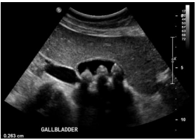
Posterior enhancement
Opposite of posterior shadowing, in which there is brightness under a low density (anechoic) object like fluid.
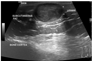
Ring down
Spaces with bubbles reverberate the liquid and create a vertical artifact.
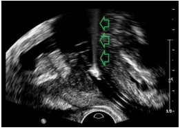
Edge
Shadow from a rounded structure. Requires scanning at a different angle.
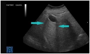
Noise
Grain. Requires decreasing the gain (amplification) or adjusting the Time gain Compensation (TCG).
Probes
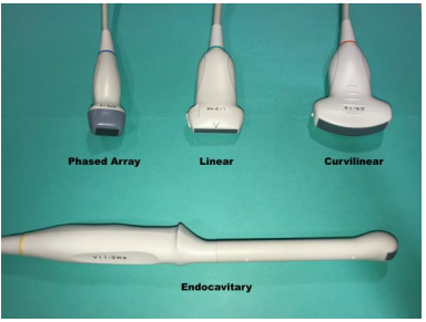
Linear (12L): Vascular exams like carotid doppler
Curvilinear (4c): Wide view angle for abdominal visualization or obstetrics
Phased array (3S): Cardiac visualization (e.g., echocardiogram)
Endocavitary probes: Body cavities
