Ectoderm - Neurulation
Development of the ectoderm
The ectoderm does not ingress through the primitive streak.
Steps
- Notochord formation
- Neural tube formation
- Notochord secretes sonic hedgehog (SHH) which controls ventralization of the neural tube
- The ectoderm secretes Wnts and BMP4 which controls dorsalization of the neural tube
- Noggin is released and inhibits BMP4/7. This negative feedback leads to differentiation in the dorsal region
- Wnts induce the dorsal neural tube to become neural crest cells
BMP 4: Induces endoderm proliferation, promotes noggin
BMP 7: Inhibits endoderm proliferation, promotes noggin
Neuroectoderm
Around day 30, the neuroectoderm begins to diffuse into layers.
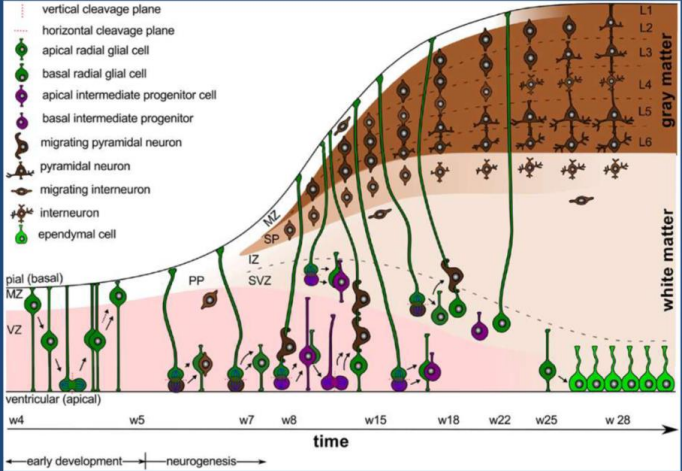
Ventricular zone: Gives rise to all neurons and glia
Intermediate zone: Derived from ventricular zone, differentiate into neurons or glioblasts
Marginal zone: Becomes white matter of spinal cord
Neural crest cells
Location of migration determines cell type. For instance, dorsal side of neural tube becomes the facial bones (only bones derived from ectoderm cells).
Spinal meninges develop from the neural crest cells and paraxial mesoderm
- Dura mater: Outer layer of meninges (hard)
- Arachnoid mater: Middle layer of meninges
- Pia mater: Inner layer of meninges (soft)
Other tissues from the neural crest cells
- Sensory neurons in the dorsal root ganglia
- Sympathetic chain ganglia
- Pigmented cells (melanocytes)
- Dentin in teeth
- Adrenal medulla
- Spiral septum (deficit in crest cells leads to persistent truncus arteriosus)
- Schwann cells and glial cells
Spinal cord
Nervous System Primer > Nerves
BMP signaling on the dorsal side results in the alar plate (afferent neurons). SHH signaling on the ventral side results in the basal plate (efferent neurons). The sulcus limitans separates the alar and basal plates.
Cerebral development
The cerebral hemispheres grow posteriorly over the brain, enveloping it.
radial glial cells → progenitor cells → neurons → glial cells
Week 5: Apical and basal radial glial cells switch from symmetric (lateral) to asymmetric (differentiation) cell division.
Week 7: Formation of the subventricular zone (SVZ) which manages the migration of pyramidal neurons

Gyrification
The process of folding within the brain. By week 10, there are secondary folds present. At week 24-26, tertiary folds are present. White and gray matter appear at the same time gyrification is occuring.
The telencephalon undergoes the most gyrification, making up the cerebrum. It will grow over the mesencephalon (pons/cerebellum), while the mesencephalon grows over the medulla.
Gyrus: Top of fold
Sulcus: Bottom of fold
Folding mechanisms
Mechanical buckling: Force of dividing neurons in confined space
Axonal tension: Pull from highly connected cortical areas cause inward folding
Differential tangential expansion (most popular): Differences in neuroprogenitor cell division rates

Clinical Pearls
Lissencephaly
Absence of folds in the brain. Overly thick gray matter development causes a smooth brain and an unmyelinated area. This does not appear until week 5.
Symptoms: Motor problems, seizure, diminished cognitive function
Treatments: Anti-seizure drugs and physical therapy
Polymicrogyria
Over gyrification. Results in increased cell death in the center of the brain.
Symptoms: Motor impairment, speech impairment, seizure, cognitive impairment
Treatments: Antiepileptic medication, general pharmacological intervention
Microcephaly
Abnormally small brain. Altered cleavage plane during progenitor cell division reduces symmetric division in favor of asymmetric division. Results in low progenitor cell counts, decreasing brain volume.
Causes: Chromosomal abnormalities, deletions, Zika virus, PKU, malnutrition
Neural tube defects
Spina bifida occulta
Failure of vertebral arch to close. Presents with a dimple and tuft of hair on back. No treatment required.
Meningocele
Sac-like protrusion along the vertebral column filled with cerebrospinal fluid and meninges. Surgery required to correct gait.
Myelomeningocele
Sac-like protrusion along the vertebral column filled with cerebrospinal fluid, meninges, nerve roots, and spinal cord. Surgical intervention is required. Paralysis is a likely outcome.
Folic acid deficiency
Can cause neural tube defects as development requires an abundance of nucleic acid for DNA synthesis and for neurons to divide timely and correctly.
Neural crest cell deficits
SOD deficiency
Causes buildup of free radicals and neural crest cells become vulnerable to alcohols and retinoic acids (vitamin A)
Autism (or the 'tism as we call it back home)
Research suggests neural crest cell defects may be a cause.
Hypoplastic left heart syndrome
In which the left ventricle and aorta are too small. Babies will often have an atrial septal defect between the atria. Norwood procedure required which shunts the aorta to the pulmonary vein.