Inflammation Images
For inflammation pathology, see here: Inflammation and Repair
Acute Inflammation
Lungs and alveolar spaces
Vasodilation, increased vascular permeability, neutrophils, vascular stasis.
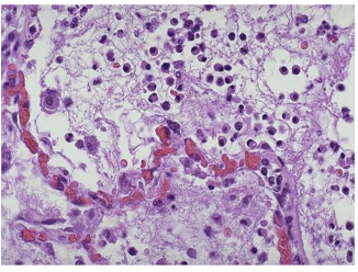
Bronchopneumonia. Neutrophil exudate seen more clearly here, resulting in productive cough.
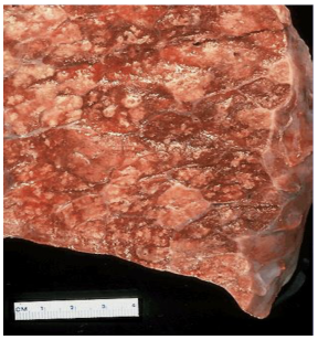
Necrotizing Pneumonia. Extensive neutrophilic exudate seen here with necrotizing pattern in center.
Mucosal surface with Ulceration of Gastric Mucosa. Epithelial tissue has sloughed off, with vasodilation of surrounding ulcer.
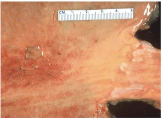
Lung surface with Necrotizing Bronchopneumonia. White splotches are necrotizing pattern.
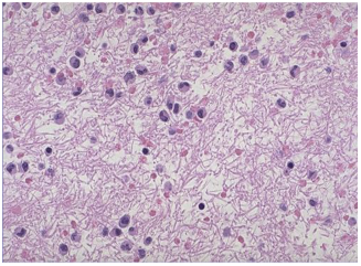
Fibrin mesh
Vasodilation, increased vascular permeability, neutrophils, vascular stasis.
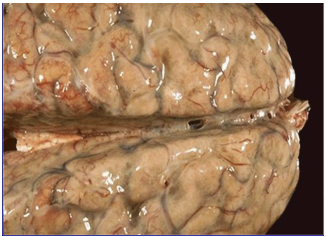
Brain
Bacterial meningitis. Red vessels from vasodilation. Pus wedge on left.
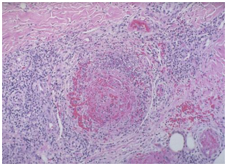
Blood vessel
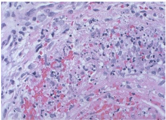
Degradation of vessel wall with coagulation (thrombus formation).
Higher magnification image.
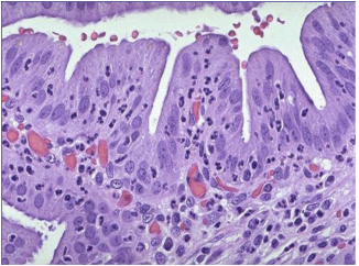
Gallbladder
Columnar epithelium. Neutrophil infiltration of mucosa/submucosa (dark spots).
Chronic Inflammation
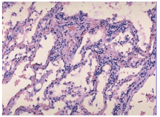
Lung
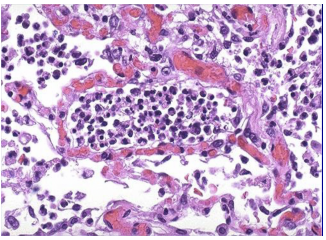
Influenza A. Interstitial infiltrate from lymphocytes creates swelling in alveolar walls.
Synovium Joint
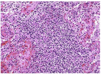
Rheumatoid arthritis. Lymphoid aggregate in dark region.
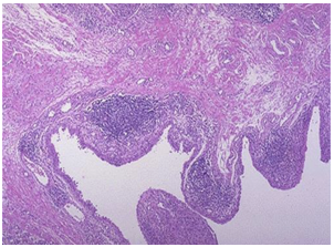
Granulomatous Inflammation
Lung
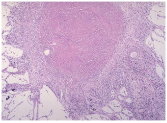
Tuberculosis with caseous necrosis.
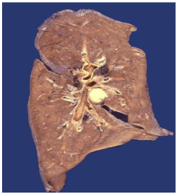
Side-by-side pulmonary granulomas.
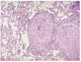
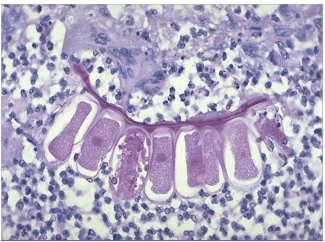
Foreign Body Giant Cell Reactions
Sutures
Dark streak center-right.
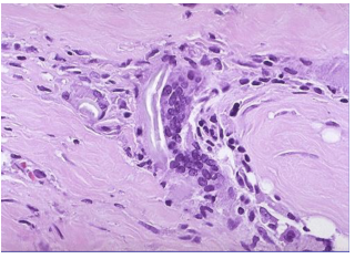
Aspirated Vegetable
Upper left of center. Neutrophils also present.
Healing and Granulation Tissue
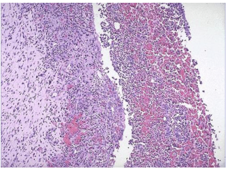
Abscess
Granulation tissue and abscess to the left. New vessels forming. Pus on right side.
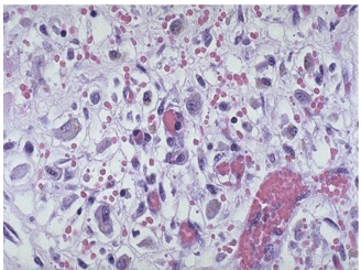
Granulation tissue
Capillaries, fibroblasts, variable amounts of inflammatory cells.
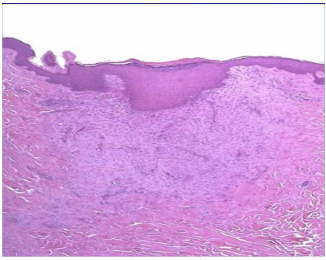
Healing biopsy site
Top layer is re-epithelialized skin. Underneath is granulation tissue, small capillaries, and fibroblasts forming collagen.
Silicone granulomas
Prominent fibrosis. Dark center is collagen.
