Necrosis And Apoptosis Images
For more detail, see Necrosis and Apoptosis Primer
Steatosis: Abnormal accumulation of fats within parenchymal cells
Lipofuscin: Pigmented granules in cells
Heterophagy: Taking up materials from the external environment
Images of Healthy Tissue
For more on the epithelium classification, see here: Histological Techniques, Epithelium, and Glands > Epithelium
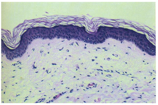
Keratinized stratified squamous epithelium
Squamous cells sitting atop a basement membrane, with flat cells on top. The apical surface has keratin.
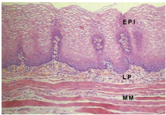
Non-keratinizing squamous epithelium
This is from the esophagus. The basal layer has higher mitotic activity than the apical layers.
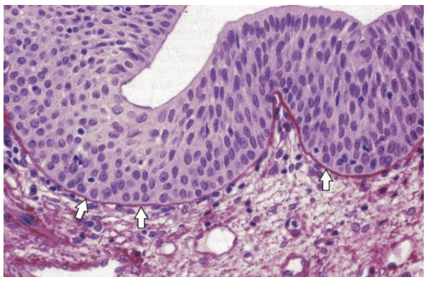
Transitional uroepithelium
From the bladder.
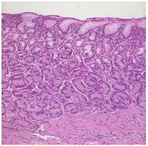
Glandular epithelium
From the stomach.
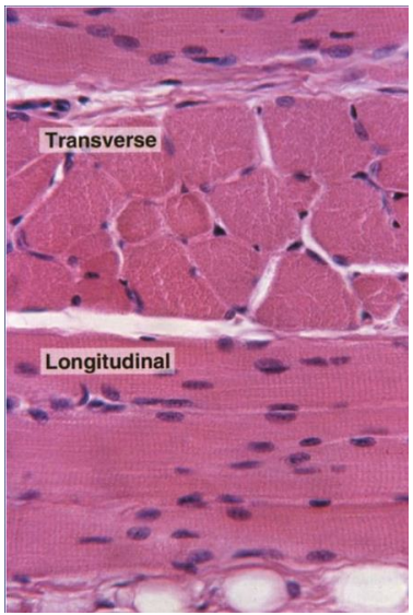
Skeletal muscle
Cross striations between fibers.
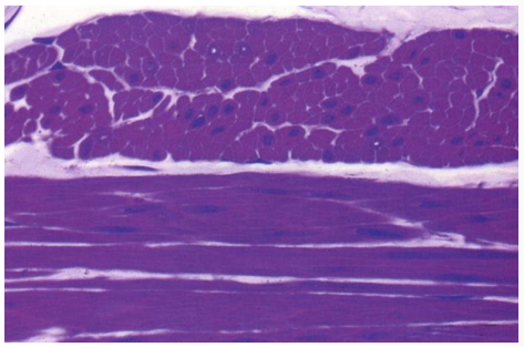
Smooth muscle
Images of Atrophied Tissue
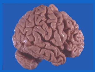
Brain
This is cerebral atrophy in a patient with Alzheimer's disease, which will lead to cognitive issues. The gyri are narrowed and sulci widened toward the front. This is most likely to be seen in regions with decreased blood supply (chronic ischemia).
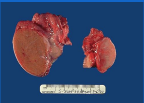
Testis
Parenchymal tissue from the testis to the right has atrophied and is smaller and more fibrotic as a result. This can be caused by vascular occlusion, hormonal imbalances, cryptorchidism (undescended testis).
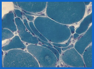
Muscle
These muscles have some cells smaller than the others. This case is caused by Denervation Atrophy (loss of nerve stimulation).
Images of Tissues with Hyperplasia
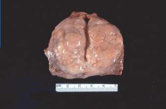
Prostate
While a normal prostate is 3-4 inches, the number of prostatic glands and stroma have increased here. This process is nodular (non-uniform) and makes it bumpy. This can be caused by Benign Prostatic Hyperplasia (BPH), which is common in older men and can cause urinary frequency/incontinence.
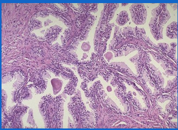
This is an example of prostate tissue which has normal looking cells, but there are simply way to many. An excess of glandular epithelium.
Images of Tissues with Metaplasia
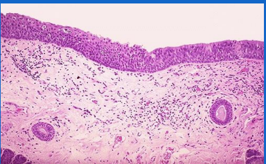
Laryngeal epithelium
The cells at the right (pseudostratified columnar) are normal, but those on the left (squamous) have been exchanged for a more resilient cell type. This is commonly found in locations of irritation (GI or respiratory tract). This tissue comes from a chronic smoker and the patient may will lose cilia in this region.
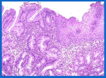
Esophageal mucosa
The cells on right (squamous) are normal, but those on the left (glandular, columnar) have been exchanged. This occurred from chronic irritation from acid reflux, and the new cells developed in order to produce mucin for protection. This presentation is called Barrett's Esophagus.
Images of Tissue with Coagulative Necrosis
Note that in the brain, ischemia causes liquefactive necrosis rather than coagulative.
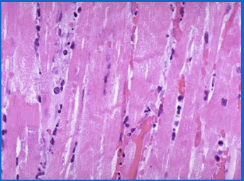
Myocardium
This nuclei of this tissue is being lost and the cells are blending together. The lost nuclei causes cells to become eosinophilic. Inflammatory cells (neutrophils) are present. This is caused by ischemia.
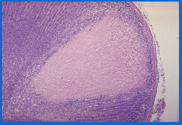
Adrenal Cortex
The pale tissue here has lost blood supply, also due to ischemia. Note the loss of nuclei.
Images of Tissue with Liquefactive Necrosis
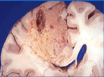
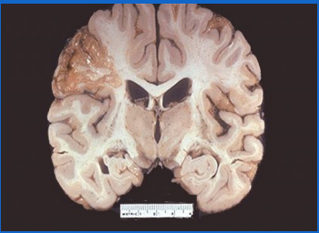
Brain
There is a loss of blood supply here in the upper left from a cerebral infarction (stroke) which has cause an abscess (collection of pus, neutrophils, dead tissue, and fibrin). The neutrophils have triggered the enzymatic digestion.
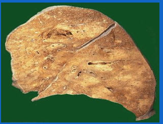
Lung
There are two abscesses in this image, on in each lobe. The white regions represent high density of neutrophils. This is a complication of severe pneumonia from virulent organisms (e.g. Staphylococcus aureus). These tend to appear in the right posterior lung.
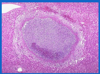
Liver
This is a small abscess filled with many neutrophils, an localized example of liquefactive necrosis.
Images of Tissue with Caseous Necrosis
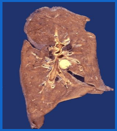
Lung
This is a hilar lymph node which has taken on a cheesy tan appearance, a result of the combination of coagulative and liquefactive necrosis in the setting of granulomatous inflammation (a subtype of chronic inflammation). A granuloma is a ball of macrophages and other inflammatory cells. In this case, the granuloma with central necrosis indicates tuberculosis.
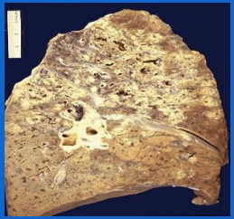
This is a more extensive example with a confluence of granulomas. The tissue destruction is so extensive there is some cavitation (cystic spaces) formed as debris drains.
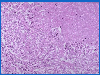
This microscopic image of lung tissue is characterized by acellular pink areas surrounded by the granuloma inflammation. The blueish/purple cells are macrophages.
Images of Tissue with Gangrenous Necrosis
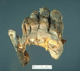
Toes
Gangrenous necrosis will feature necrosis of many body tissues, generally caused by ischemia. This is a "dry" gangrene from frostbite.
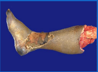
Lower limb
This necrosis also resulted from a loss of blood supply, but is a "wet" variant in which there is a present liquefactive component. This case is a symptom of diabetes which reduces vascularity and wound healing.
Images of Tissue with Accumulations
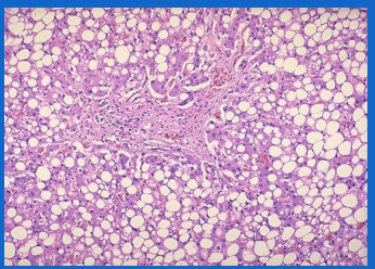
Liver
Here, excess fats have been stored (steatosis) as a result of reduced lipoprotein transport from chronic alcoholism.
Hilar lymph node
This features accumulation of anthracotic pigment (anthracosis), a carbon pigment. This case is from a chronic smoker, but the pigment itself does not actually cause dysfunction.
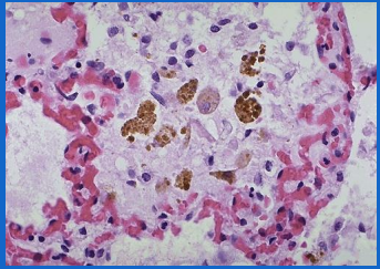
Lung
This alveolar tissue has collected brown hemosiderin from macrophages, a side effect of the breakdown of red blood cells (releasing heme).
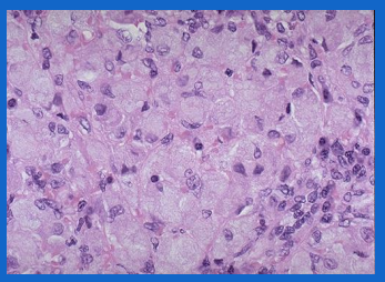
Spleen
The large pale cells contain accumulated storage from lack of an enzyme. This example is from Gaucher's disease.
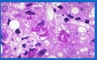
Liver
These red globular materials are Mallory Bodies damaged by chronic alcoholism.
Images of Tissue with Calcification
Stomach
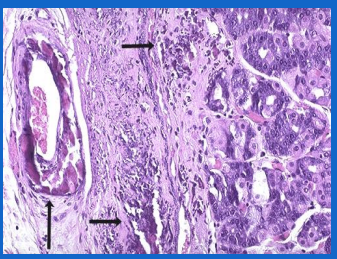
The far left demonstrates an artery with calcification of its walls, as well as an irregular blue/purple deposit in the submucosa. Calcified specimens feel firm and gritty as well as basophilic in appearance.
