Histological Techniques, Epithelium, And Glands
Microscopy
| Type | Mechanism | Resolves |
|---|---|---|
| Light microscope (LM) | Glass lens | Large structures (primarily nucleus) |
| Transmission electron microscope (TEM) | Magnetically focused electron beam | Small cell organelles, used sometimes for freeze fracture |
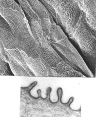
| Scanning electron microscope (SEM) | Electricity scanning (non-penetrating) | Surface topography |
Freeze fracture: Tissue is frozen and split into inner and outer leaflets. Can be used to see proteins embedded in the membrane.
Preparation
Preparation steps:
- Fix the tissue to stabilize it
- Dehydrate
- Embed it into a hardening material
- Section into thin slices
- Mount on slide and stain
| Type | Fixation | Dehydration | Embedding | Sectioning | Mounting/Staining |
|---|---|---|---|---|---|
| LM | Formalin | Ethanol/xylene | Paraffin wax | Steel blade/microtime | See below |
| TEM | Formalin, glutaraldehyde, osmium, tetroxide | Ethanol | Epoxy or epon resin | Diamond knife | Lead citrate and uranyl acetate |
| SEM | Formalin | Ethanol | Gold | NA | NA |
Common stains
Hematoxylin and Eosin
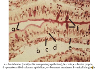
Hematoxylin stain nucleus blue, eosin stains cytoplasm pink
Silver
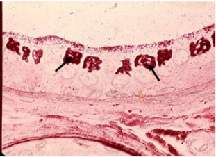
Stains golgi (below), reticular fibers (type III collagen)
Other stains
- Periodic acid schiff: Mitochondria, glycogen, glycoproteins stained red
- Aldehyde fuchsin: Elastic fibers stain red/deep purple
- Orcein: Elastic fibers, collagen fibers, cytoplasmic counterstain stain brown
Epithelium
How to identify epithelium:
- Found on all body surfaces
- Covers all passage which connect to exterior
- Makes up secretory cells of glands
- No blood supply (avascular)
- Lies on basement membrane or basal lamina
Epithelium is either glandular or surface. It is classified by:
- The number of cells: Simple or stratified. Simple is when cells rest directly on basement membrane. Stratified is stacked layers.
- The height/shape of the surface layer: Squamous, cuboidal, or columnar. Squamous is flat (squat).
Other layer types:
- Pseudostratified: Not stratified, one layer but looks like several because nuclei are in different parts of the cell
- Transitional: Multiple variations in one tissue
Note: if a cell is binucleated and stratified, it is BLADDER tissue
| Type | Diagram | Locations |
|---|---|---|
| Simple squamous | 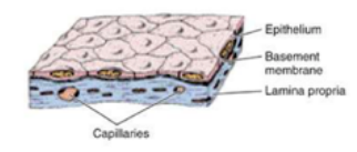 | Endothelium, mesothelium, parietal layer of Bowman's capsule, thin segment of loop of Henle, rete testis, pulmonary alveoli |
| Simple cuboidal | 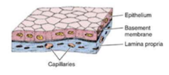 | Thyroid, choroid plexus, ducts of glands, inner surface of lens, covering of ovary |
| Simple columnar | 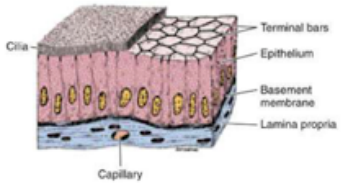 | Surface stomach, small/large intestine, gallbladder, excretory ducts of glands, uterus oviducts, small bronchi of lungs |
| Stratified squamous | 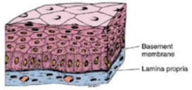 | Tympanic cavity, buccal surface, esophagus, epiglottis, conjunctiva, cornea, vagina, skin epidermis, gingiva, hard palate |
| Stratified columnal | 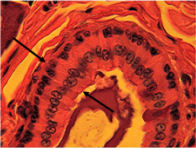 | Male urethra, fornix of conjunctiva, large duct execretory glands |
| Stratified cuboidal | 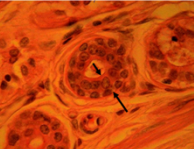 | Sweat ducts |
| Pseudostratified | 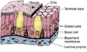 | Large excretory ducts of glands, parts of male urethra, epididymis, trachea, bronchi, eustachian tube |
| Transitional | 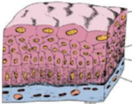 | Bladders, ureters, prostatic urethra |
Glands
Definitions
Glandular epithelia are groups of cells which secrete things. Exocrine glands have an acinus (secretory portion) and a duct (conducting portion).
- Parenchyma: secretory cells
- Strome: Connective tissue between parenchyma
- Exocrine glands: Surface or cavity secretions via ducts
- Endocrine glands: Blood stream secretion
Modes of Secretion
- Merocrine (most common): Vesicular release
- Apocrine: When vesicles are so large, a portion of the cell is released (e.g. mammary glands)
- Holocrine: Whole cell is shed (e.g. sebaceous glands)
Classification
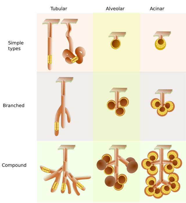
- Number of ducts
- Simple (one)
- Compound (multiple)
- Shape of secretory portion
- Tubular
- Branched tubular
- Acinar (grape shaped)
- Branched acinar
- Tubuloacinar
- Secretion type
- Serous (aqueous)
- Mucous (mucinous glycoproteins)
Cell surface specializations
Microvilli
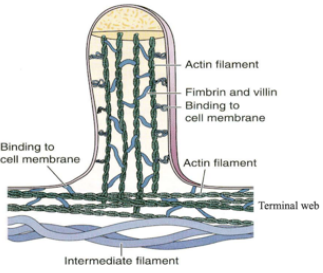
Filled with actin connected to terminal web, surrounded by glycocalyx containing glycosylated products
Cilia
More brush like or fibrous than microvilli. Made of 9 + 2 microtubule axoneme structures. Embedded in basal bodies, which are centrioles that nucleate axonemes of cilia
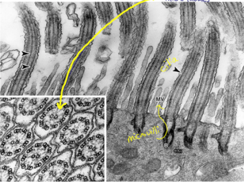
An example of an axoneme
Stereocilia
Very thin microvilli found in male reproductive tract (epididymis) and vestibular apparatus of inner ear.
Microplicae
Folds that can appear on the surface of cells, especially the GI
