Hematopoeisis
Fetal Hematopoiesis
First blood cells formed in primordial phase in the [[Fertilization and Implantation#4. Yolk sacs (12 - 13 days)|secondary yolk sac]]. In the second trimester, the liver and spleen begin to produce blood cells in the hepato-spleno-thymic phase in which precursor granulocytes, megakaryocytes, and erythroblasts are created. This phase peaks 5-6 months post-fertilization.
The final hematopoiesis phase is the medullo-lymphatic phase which occurs in the bone marrow and lymph nodes. This occurs around 4 months and continues into the 3rd trimester.
Ontology follows phylogeny: The progression of hematopoiesis in fetal development closely resembles the phylogenetic progression of lymphoid tissues in vertebrates.
Non-Encapsulated Lymphoid Tissue → Encapsulated Lymphoid Tissue (Thymus & Spleen) → Bone Marrow
Post-partum/Adult Hematopoiesis
Occurs in bone marrow of axial skeleton and distal long bones. As we age, hematopoiesis in long bones declines in favor of the axial skeleton. Over time, the yellow marrow increases relative to red marrow, which decreases our ability to perform hematopoiesis as efficiently. This is a contributor to immune system decline with age.
If bone marrow cannot produce blood cells, extramedullary hematopoiesis can occur. Will occur in tissues involved in fetal hematopoiesis.
When performing a biopsy for bone marrow analysis
- Adults: Sternum and sinuous process of vertebrae
- Infants: Shaft of tibia
Blood Lineages
Hemocytoblast is the pluripotent stem cell for all blood cells.
Hematology > Blood composition
Myeloid cells
- Erythrocytes
- Megakaryocytes (platelets)
- Granulocytes
- Monocytes
Lymphoid cells
- B lymphocytes
- T lymphocytes
Bone marrow components
- Nutrient Arteriole: provides nutrients necessary for the development of blood cells
- Stromal Cells: reside under the epithelial lining; produce hematopoietic short-range regulatory molecules and maintain the hematopoietic inductive environment
- Macrophages: usually found near sites of erythropoiesis in erythroblastic islands; engulf nuclei that have been expelled from orthochromatic erythroblasts (normoblasts)
- Endothelial Cells: the lining of blood vessels between which developing blood cells escape into the sinusoidal lumen
Blood Cell Production
Erythropoiesis
Erythropoietin (EPO) stimulates erythrocyte production. It is produced by the kidney in response to low blood oxygen detected by renal interstitial peritubular cells. Some EPO is also produced by the liver.
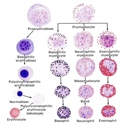
Identification of erythrocyte stages
- Proerythroblast: Has lots of RNA in cytoplasm
- Basophilic erythroblast: RNA present and stains BLUE
- Polychromatophilic erythroblast: In the sast stage in which chromatin is not condensed (mitosis can occur). Hemoglobin production begins and will color cell pink
- Normoblast (Orthochromatophilic Erythroblast): Condensed nucleus, no DNA accessible for DNA replication
- Reticulocyte (1% of RBCs): No nucleus present, but as some RNA which will stain BLUE from cresyl blue supravital stains
- Mature erythrocyte: No RNA, biconcave disk
Granulopoiesis
Identification of granulocyte stages
- Myeloblast: No granules
- Promyelocyte: Non-specific granules, oval nucleus, golgi complex
- Myelocyte: Has specific granules (basophilic, neutrophilic, or eosinophilic), oval nucleus
- Metamyelocyte: Has kidney bean nucleus, atrophic golgi complex, amoeboid movement
- Band cell: Horseshoe-shaped nucleus
- Mature granulocyte: Segmented nucleus

Monopoiesis
Macrophages are named for their tissues. When a monocyte finished developing and leaves the bloodstream, it is considered a macrophage.
Monoblast → Promonocyte → Monocyte → Tissue Macrophage
- Liver: Kupffer Cell
- Bone: Osteoclast
- Brain: Microglial Cell
- Epidermis: Langerhans Cell
Thrombopoiesis
Induced by thrombopoietin (TPO)
- Megakaryoblast (2n-64n): Multiple replications of DNA but no cell division
- Reserve Megakaryocyte (64n): No shedding of cytoplasmic blebs as platelets
- Platelet-Forming Megakaryocyte (64n): Visibly shedding platelets into the sinusoids from the abluminal surface
Classification of Leukemias
A lymphoma is a neoplasm of lymphocytes. A leukemia is a neoplasm of leukocytes.
Hodgkin's Lymphoma: Characterized by presence of Reed Sternberg cells. Has 5 subtypes
Non-Hodgkin's Lymphoma: 12 B cell and 12 T cell subtypes
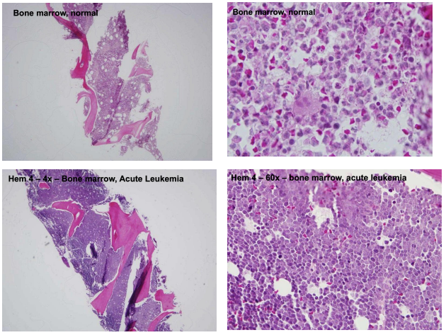
Acute leukemia: Cancer of immature blood cells. Deficiency in mature forms of these cells will exacerbate the condition
Chronic leukemia: Cancer of mature blood cells (less aggressive than acute)
