Cells And Organelles Primer
Histopathology
Use of microscopy to determine manifestation of disease.
Concentration of Organelles
Cell types will have various concentration and sizes of organelles.
In polarized cells:
- Apical surface: will typically face a lumen
- Basolateral surfaces: For cell to cell communication
Non-polarized cell are generally under differentiation.
Plasma membrane
See: Lipids, Membranes, and Membrane Proteins
Phospholipid bilayer: Two long chain fatty acids, three carbon glycerol backbone. Fatty acids are hydrophobic, glycerol is hydrophilic.
Intercellular junctions
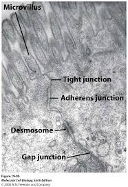
Tight junctions
Occur close to luminal surfaces (apical surface). Are permeability barriers (e.g. blood brain barrier). The membranes appear adhered to each other at various points.
Anchoring junctions
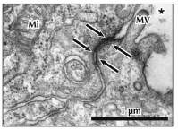
Occur at lateral borders. Resist stress
Types:
- Zonula adherens which are anchored by actin.
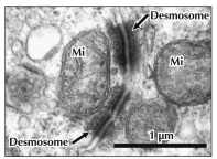
- Desmosomes (macula adherens) which are anchored by intermediate filaments. (e.g. plaques in plasma membrane, skin)
Nucleus
- Stores DNA
- Transcription factors turn genes on and off
- RNA and DNA and polymerases are enzymes which make DNA and RNA copies
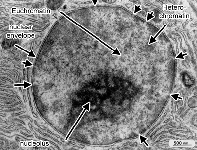
- Nucleolus: Sub-nucleus, ribosome synthesis takes place here
Note on PCR Testing
Convert RNA to DNA and then replicate (amplify) DNA. To separate DNA strands, must be heated. A taq polymerase from an organism adapted to higher temperatures is used for this case.
Nuclear envelope
Double membrane formed in the rough endoplasmic reticulum (rough ER). Has an inner nuclear membrane with specialized proteins that interact with lamina and chromatin.
Nuclear pore complexes allow nuclear proteins and ribosomal proteins into the nucleus and allows mRNAs/ribosomal subunits out of the nucleus
Ribosomes
Composed of RNA and associated proteins, very electron dense. In inactive form will appear as a single ribosome in cytoplasm. In active form will group together in a "rosette" polyribosome structure along a thread of mRNA. Subunits and proteins synthesized in the nucleolus and enter the cytoplasm via nuclear pores. Highly basophilic.
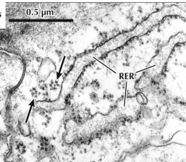
Large subunit: 2 RNA / 49 proteins
Small subunit: 1 RNA / 22 proteins
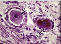
Nissl body: High protein synthesis specifically in nerve cells
Endoplasmic reticulum
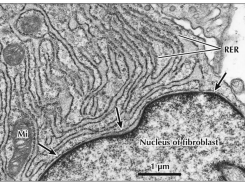
Composed of tubules, circular vesicles, and flattened membranous sacs (cisternae).
Rough ER
Continuous with nucleus. Membrane studded with ribosomes. Network of cisternae and vesicles.
Functions:
- Protein translocation
- Protein folding and oligomerization
- Carbohydrate addition
- Protein degradation/banishment

Smooth ER
Membrane is smooth (no ribosomes). Network of tubules and cisternae. Its function will depend on body location.
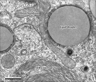
Body locations:
- Liver (hepatocytes): Carb metabolism
- Steroid secreting cells (ovary, testis, adrenal): Lipid/lipoprotein synthesis
- Muscles: Calcium regulation
Functions:
- Detoxification
- Lipid metabolism
- Heme metabolism
- Calcium release
ER storage diseases
Unfolded protein response (UPR) results from prolonged or acute stress. Activates pathway to kick out unfolded proteins. Has been implicated in diabetes, Parkinson's, atherosclerosis.
Protein misfolding
Cystic fibrosis transmembrane conductance regulator (CFTR): Regulates movement of ions and water into and out of the cell.
When a CFTR protein is misfolded, can no longer be placed in apical surface of the cell by COP II. Therefore, it will build up in ER and trigger UPR and accumulate thickened mucus in airway.
Transport out of ER
Organelle Function > Endocytosis and exocytosis
Three modes of transport:
- Bulk flow: Bud off entire potions of membrane into cytoplasm (usually headed to Golgi apparatus). Does not interact with export signals.
- Partitioning with the lipid bilayer
- Signal-mediated sorting: Interact with COP II to concentrate products into carriers
Three coat proteins:
COPII: Coat vesicles from the ER.
COPI: Coat vesicles from cis/medial regions of golgi
Clatherin: Coats vesicles from trans golgi, endosome, and plasma membrane
Golgi Complex (Apparatus)
Located near nucleus/centrosome. Important for microtubule formation as well as transport. Vesicles pass from cis side → medial golgi → Trans golgi → Trans golgi network.
Has two sides:
- Cis Golgi: Convex and has vesicles and medial compartment (stacks of flattened saccules)
- Trans Golgi: Concave and has vesicles and vacuoles for distribution and sorting of secretory products
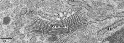
Function
- Produce (with ER) lipids
- Produce lysosomes
- Post translational modifications
- Sort/send secretory proteins from RER
Golgi in Alzheimer's
Neurons in the basal forebrain and hippocampus have some fragmentation/atrophy of golgi. The golgi loses cis/trans polarization and form small vacuoles which distend into proximal dendrites. This breaks the neuron.
Lysosomes
Membrane-bound vesicles and vacuoles derived from trans golgi complex vesicles. Abundant in phagocytosis cells. Optimal enzymatic pH is 4-5.
Functions:
- Eats viruses, bacteria, and other pathogens
- Removes worn organelles
- Aid in autolysis of cells as lysosomes explode and release their enzymes
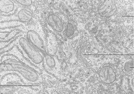
Lysosome stages
- Primary: Electron dense, homogenous
- Secondary: Larger, heterogenous looking, have some digested material
- Tertiary: Beat the fuck up looking. Full of debris.
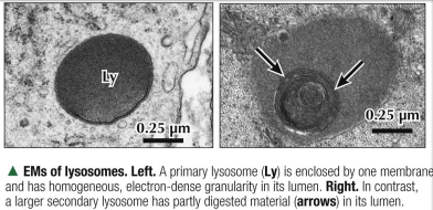
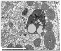
Clinical Pearl: Tay sachs
Lysosomal storage problem, rapidly fatal. Hexosaminidase A (hex A) is deficient which allows ganglioside GM2 to accumulate in cells which is no bueno.
