Organelle Function
Cytoskeleton
Protein structures which provide structural integrity.
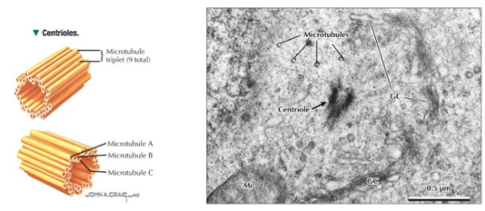
Microtubules
Hollow, unbranched, and cylindrical. Are polar (+/- ends) with variable length. When organized into triplets, they are referred to as basal bodies.
Functions:
- Movement of flagella and cilia
- Cytokinesis
- Transport of organelles (kinesin moves towards +, dynein moves towards -)
- Buddies with the Golgi, holding it in place and acting as its transport network
Location: Neurons, platelets, leukocytes, any dividing cell
Neurons have a lot of microtubules because they are constantly sending neurotransmitters
Actin filaments
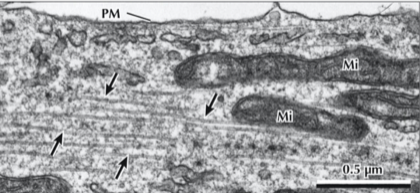
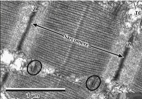
Flexible, thin rods. Thin filaments are actin, thick filaments are myosin.
Functions:
- Resist cell shape change
- Muscle cell contraction (sarcomere has thick/thin filaments)
- Cell movement, cytokinesis, phagocytosis
Location: All cells
Intermediate filaments (IF)
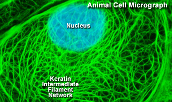
Wavy, unordered bundles of thin rods
Functions:
- Mechanical support/structural integrity
- Support desmosomes
- Link nucleus to plasma membrane
- Reinforce nuclear envelope
Location: Cells with a lot of mechanical stress
Types:
- Nuclear lamins: Critical for scaffolding the inner nuclear envelope and organizing DNA in interphase
- Desmosomes: Helps transmit forces between cells
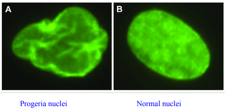
Clinical peal: Progeria (premature aging)
Causes by a mutation resulting in the production of Progerin instead of normal Lamin A which provides the nucleus structural integrity. As a result, we see "blebbed" nuclei which ages the body rapidly. Short life expectancy.
Endocytosis and exocytosis
Process by which cells send and receive material
Endocytosis
Cells and Organelles Primer > Transport out of ER
Stuff coming in the cell, via invagination of the cell membrane
Types:
- Fluid-phase (pinocytosis): Non-selective, triggered by difference in gradient
- Receptor-mediated: Selective (e.g. by a hormone or growth factor). Uses clatherin coated vesicles
- Caveolae: Mediates transcytosis using caveolin coated vesicles
- Intakes at one end of cell and exists at other end
- For example, if a cell wants to push bacteria into the lymph
Exocytosis
Transport out of the cell. Cells often branch off the golgi and then fuse with the plasma membrane to release the contents.
For more see: Histological Techniques, Epithelium, and Glands > Modes of Secretion
Peroxisomes
Have a single thin membrane with a granular matrix on the inside. They uptake proteins from the cytoplasm to self-replicate.
Functions:
- Oxidation
- Respiration
- Fatty acid (long-chain) metabolism
- Regulation of hydrogen peroxide (H2O2)
- Bile acid metabolism
Location: Liver cells and kidney tubules, often near RER
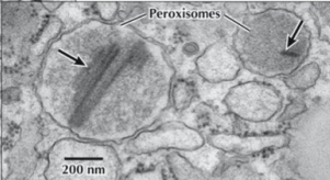
Clinical pearl: Zellweger syndrome (cerebrohepatorenal syndrome)
A mutation prevents peroxisomes from importing proteins across their membrane (unable to replicate). This causes brain, kidney, and liver abnormalities. Because peroxisomes synthesize plasmalogens (the predominant phospholipid in nervous tissue myelin) deficient myelination of neural cells.
Inclusions
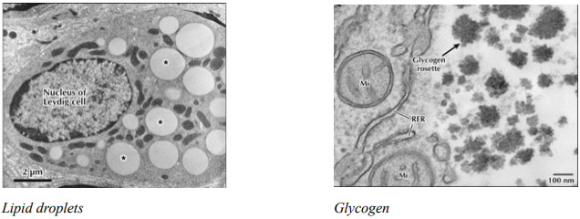
Subdomain of cellular content which is generally transient. Includes glycogen, lipid droplets, and pigment granules.
Glycogen: Found in muscle or liver cells near the endoplasmic reticulum (where glycogenolysis may occur). Is dark and forms clusters called rosettes in hepatocytes (liver).
Lipid droplets: Fat globs inside cells, appearing as white or light circles.
Mitochondria
Typically scatter through cell but can concentrate if there is a lot of energy use. When there is mitochondrial myopathy, it typically affects the muscles first, since these cell types use a lot of energy, all the time.
The only place you will not find mitochondria is mature erythrocytes.
Structure
- Outer membrane: Porin allows for passage of molecules
- Intermembrane space: Between outer membrane and inner membrane. ATP synthesis and apoptosis
- Inner membrane: Encloses the matrix with folds called cristae (increasing the surface area)
- Matrix: Has enzymes for TCA/Krebs/Citric acid cycle. Hosts mitochondrial DNA/ribosomes
Clinical pearl: Wilson disease
Mitochondria will have dark, dilated cristae and a non-uniform look
Centrioles
Structure: Made up of 9 microtubule triplets
Function: Helps transport Golgi vesicles to other parts of the cell
Location: Varies based on cell stage. Will migrate to poles during mitosis.
Centrosome
Structure: Made up of a pair of centrioles
Function: Organizes microtubules and is the origin of new microtubules/the mitotic spindle
Location: Near the nucleus, associated with the Golgi
