Connective Tissue
Classification (Type)
Proper
- Loose
- Dense
- Regular
- Irregular
Supporting
- Adipose tissue
- Elastic tissue
- Mucus tissue
- Hematopoietic
Special properties
- Cartilage
- Bone
Classification (Origin)
Hematopoietic
For more, see here: Hematopoeisis
- Red blood cells
- White blood cells
- T/B cells
- Mast cells
- Microglia
- Langerhans cells
- Osteoclasts
Undifferentiated mesenchymal
- Fibroblasts
- Adipocytes
- Chondrocytes
- Osteoblasts
- Mesothelial cells
- Endothelial cells
- Smooth muscle cells
Cellular Components
Fibroblasts
Structural, produce fibers and ground substance.

Adipose cells
Unilocular adipocytes
Function: Fat cells, energy storage.
Features: Large fat cells which include one lipid droplet. Lots of open space with a thin cytoplasm along edge.
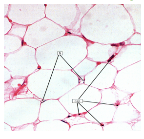
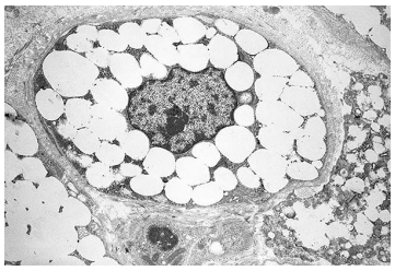
Multilocular adipocytes
Function: Only present in neonates as a form of heat generation
Features: Include multiple lipid droplets and mitochondria
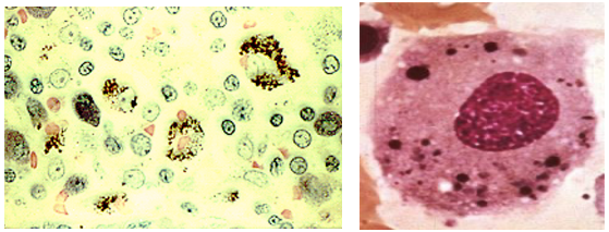
Macrophages
Function: Phagocytic cells which take up debris
Features: Visible vesicles
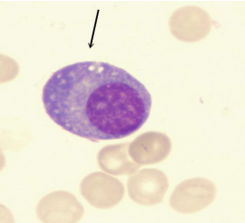
Plasma cells
Function: Derived from B cells, produce immunoglobulins.
Features: Eccentric nucleus, cartwheel arrangement of heterochromatin, abundant RER.
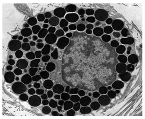
Mast cells
Function: Basophilic molecules containing pharmacologically active substances mediating allergy response. IgE on surface trigger an allergy response (heparin, histamine, leukotrienes, proteoglycans, ECF-A).
Features: Large basophilic granules around a nucleus. Stains dark.
Amorphous tissue
Known as ground substance. Molecules composed of varied protein and sugar ratios.
Glycosaminoglycans
For more, see here: Carbohydrates and Sugar Code Primer > Glycosaminoglycans
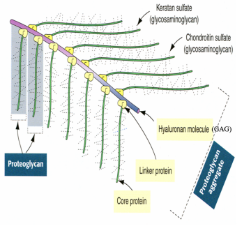
Disaccharide sugars containing amino sugars. Hold water. An example is hyaluronic acid (in eye).
Proteoglycans
A core protein connected to glycosaminoglycan chains. Brush like.
Glycoproteins
For more, see here: Protein Primer > Glycoproteins
Globular proteins with branched sugar residues found in basement membrane. Binding sites for cells, collagen, and heparan. Examples include fibronectin, laminin, and integrins.
Fibrous components
Collagen fibers
For more, see here: Protein Primer > Collagen. Seen as thick fibers with striations.
Functions
- Adhesion
- Skeletal (bones, cartilage)
- Protective (skin)
- Messenger
Synthesis
- Pre-procollagen translated from mRNA
- Procollagen formed after cleavage of signal peptide
- Hydroxylation/glycosylation (using Vitamin C)
- Alignment of chains to form triple helix
- Packaging into secretory vesicles, secretion
- Formation of tropocollagen
- Bundling to form fibril → fiber → fiber bundle
Clinical pearls
Scurvy: Resulting from Vitamin C deficiency
Ehlers-Danlos (VI): Faulty hydroxylation
Ehlers-Danlos (VII): Decreased peptidase activity (step 6)
Elastic fibers
Formed from oxytalan (fibrillin) and elastin. Oxytalan resists tension while elastin provides flexibility. Appears thin and brownish-red (stains with orecin).
Clinical pearl: Marfan Syndrome
Inherited mutation in fibrillin causing a weakened aorta, potentially leading to aortic dissection.
Reticular fibers (type III collagen)
Found in lymphoid organs and tissues. Forms scaffolding allowing lymphocytes to take refuge as the mighty fluid current rushes past. Turns black with silver stains.