Dermatologic Neoplastic Diseases
Skin Cancer Risk
The Fitzpatrick Scale assesses vulnerability to UV based on skin phenotype. Scored on a scale of I (always burns, does not tan) to VI (never burns, tans easily).
Basal Cell Carcinoma
Non-melanoma skin cancer. Most common form of skin cancer. Derived from undifferentiated pluripotent epithelial stem cells and characterized by indolent behavior (can linger for years) and unlikeliness to metastasize.

Subtypes
- Nodular: Often presents on the face. Characterized by shiny, pearly papule/nodule often with arborizing (branching) vessels.
- Superficial: Often presents on the trunk and extremities. Characterized by well-circumscribed red thin papule or plaque, sometimes with subtle rolled border.
- Morpheaform (or sclerosing): Characterized by more aggressive local destruction and appears similar to scar tissue with with white to pink atrophic plaque (fibrotic) and ill-defined borders.
Risk factors
- Intermittent intense sun exposure
- History of prior Basal Cell Carcinoma (44% relapse)
Clinical features
80% present on the head and neck.
Histology
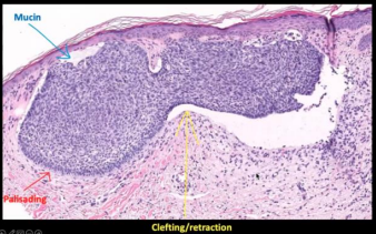
- Proliferation of blue (Basaloid) cells often connected to epidermis
- Arranged in lobules set in fibromucinous stroma
- Clefting at the periphery of tumor lobules
- Peripheral palisading

Treatment
Varies based on subtype and location.
Mohs surgery for high risk and cosmetically sensitive locations. Options for low risk variants include cryosurgery, electrodessication, topical 5-fluorouracil, or simple excision.
Hedgehog inhibitors (vismodegib) can help stop growth in advanced disease.
Pathogenesis
PTCH1 (tumor suppressor gene) inhibits smoothened (SMO) which is a regulator of cell growth.
Sonic Hedgehog (SHH) → inhibits PTCH1 → activates SMO → upregulation of GLI (transcription factor)
Dysfunctional PTCH1 means that SMO remains active even without SHH.
Clinical pearl: Gorlin syndrome
An autosomal dominant disorder for the PTCH1 gene. Characterized by lots of Basal Cell Carcinomas at an early age. Treated with an SMO inhibitor (vismodegib).
Squamous Cell Carcinoma
Non-melanoma skin cancer. Most common in African Americans.
Risk factors
- Chronic inflammation and wounds (Marjolin)
- UV exposure
- Smoking
- HPV
- Immune suppression
Clinical features
Lips, ears and anogenital regions are high-risk sites.
Actinic keratosis is the abnormal partial thickness of the basal layer and is the precursor to Squamous Cell Carcinoma. It is directly related to UV damage. It presents as a rough, erythematous papule with overlying scale.
Squamous cell carcinoma in situ (SCCIS)
Can arise from actinic keratosis or de novo, in which case it is called Bowen’s disease. It can also arise from HPV-16/18 in genital area or Erythroplasia of Queyrat (which sounds like something from Dune but it's actually penile cancer).
It presents with scaly red papule or plaque.
Invasive squamous cell carcinoma
Clinical presentation can vary. Usually presents on sun-exposed sites as a well-circumscribed nodule with central keratinaceous core. It has rapid onset (weeks) then gradual involution (months).
Associated with Muir-Torre syndrome (Lynch syndrome) and BRAF inhibitors. Must be excised.
Verrucous carcinoma
Low grade variant of Squamous Cell Carcinoma with a low risk of metastasis but high risk of recurrence. Can present on extremities (and lead to osteomyelitis) or in genital area.
Associated with HPV infection. Treatment requires excision (or amputation if severe enough).
Histology
Arranged in squamous lobules disconnected from the epidermis. Keratin pearls are a good indication of Squamous Cell Carcinoma.
Pathogenesis
Progresses from Actinic keratosis to Cutaneous Squamous Cell Carcinoma.
UVB exposure dysregulates P53, resulting in impaired apoptosis and cell proliferation.
Melanoma
Associated with sun exposure in general. Treated with excision and/or immunotherapy.
Subtypes
- Superficial: Most common type in light skin. Found on the back (for men), or legs (for women). Growth starts with slow radial phase and progresses to a rapid vertical phase.
- Nodular: Found on the trunk, head and neck. Slow growing vertical phase.
- Lentigo maligna (LM): Found on the face (nose and cheeks). Slow growing.
- Acral lentiginous: Most common type in dark skin. Found on palms, soles, or nails. Usually diagnosed at advanced stage.
Pathogenesis
Key mutations
- MAP kinase pathway (BRAF, NRAS, KIT)
- CDKN2A (tumor suppressor)
- MC1R (pigmentation gene)
ABCDE Assessment
Self-assessment tool
- Asymmetry
- Border
- Color
- Diameter
- Evolving
Histology
Pagetoid scatter (melanocytes moving up to the granular layer) is the key feature. The Breslow method assesses depth of melanoma invasion and is the best prognostic factor.