Neoplasia Images
For more detail on neoplasia, see here: Neoplasia Primer
Dysplasia
Mild
Ectocervix
Covered in squamous epithelium. Normal cervical squamous epithelial cells on the left merge into dysplastic squamous epithelium on the right. The abnormal cells on the right are disordered with almost indistinguishable cell borders, have a high nuclear to cytoplasm ratio, and have large, irregularly shaped nuclei.
Severe
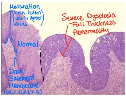
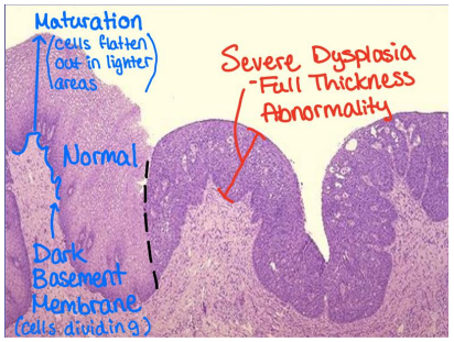
Ectocervix
Covered in squamous epithelium. Normal cells on the left show mitotically active cells near the basement membrane, causing it’s darker appearance. Normally, as cells undergo maturation, the flatten out and become lighter in appearance. Abnormal dysplastic squamous epithelial cells on the right have a high nuclear to cytoplasm ratio. Large, hyperchromatic nuclei (making them appear dark) and irregularly shaped nuclear membranes.
This case is HPV.
Neoplasm
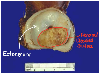
Ectocervix
The white, tortellini shape is normal squamous epithelium while the abnormal ulcerated mass circled in red is a neoplasm.
Signs and Symptoms include local bleeding, abnormal discharge, pain with sexual activity
This case is Invasive Squamous Cell Carcinoma.
Invasive Squamous Cell Carcinoma
Lamina Propria below the Basement Membrane.
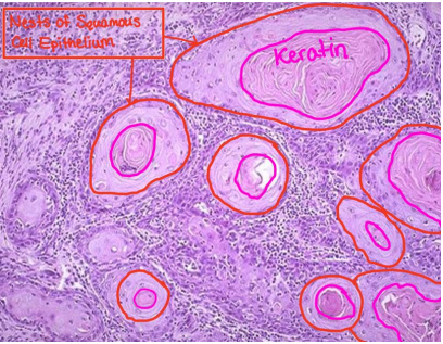
The tumor cells have grown beyond the basement membrane and invaded the submucosa (Neoplasm). Flattened squamous epithelial cells are present in large nests. These cells are well differentiated compared to their surroundings. The pink “stringy” material in the center of the nests is keratin, which is a typical protein produced by epithelial cells.
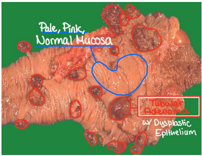
Tubular Adenomas with Dysplastic Epithelium
Cecum of the Large Intestine
The pale pink tissue is the normal mucosa. There are multiple nonspecific polyps (abnormal growths in the lumen), but their appearance suggest anything regarding malignancy. With testing, they are identified as benign neoplasms of glandular tissue and, therefore, adenomas.
This case is familial polyposis
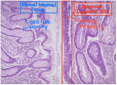
Dysplastic Epithelial Tissues
Large Intestine
The left side of the image depicts normal columnar epithelium of the intestinal mucosa. In this abnormal, dysplastic epithelium, hyperchromasia can be seen from the enlarged nuclei, a loss of polarity, and less structured cell shape.
Benign Tumors
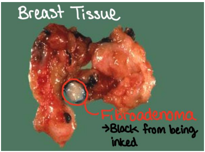
Fibroadenoma
Breast Tissue
Small benign neoplasm classified as a fibroadenoma. It is is encapsulated and well circumscribed. The lesion has a black outline from being inked. More commonly found in younger women of reproductive age.
Note: This tumor would feel soft and mobile on palpation, in contrast to most other cancers which have desmoplasia (hard and immobile).
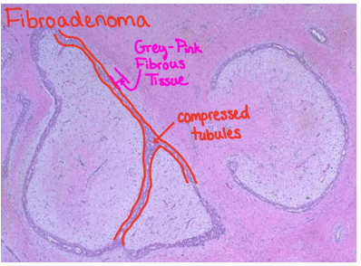
Same tumor with higher magnification. A grey-pink fibrous stroma (resulting from increased collagen) can be observed compressing the tubular ducts. Normal breast tissue is made up of clusters of tubules to form lobules.
These lesions are most likely to be found as a "breast lump" on examination of young women. They are discreet, firm, rubbery masses that are freely movable.
Leiomyomas
Uterus
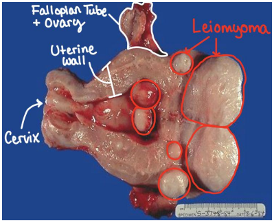
This image shows multiple benign neoplasms or different sizes. The uterus is made up of smooth muscle, hence the leiomyoma. There are defined borders of the tumors, characteristic of being well-circumscribed.
Hepatocellular Adenoma
Cut surface of Liver
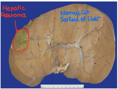
Image shows a well circumscribed hepatic adenoma. Liver doesn’t have glands, but “adeno” is used because of the increase of fibrous tissue (i.e. collagen) which is derived from the epithelium. These tumors closely mimic hepatocytes and the greenish hue is most likely from bile.
This case is exogenous steroid use.
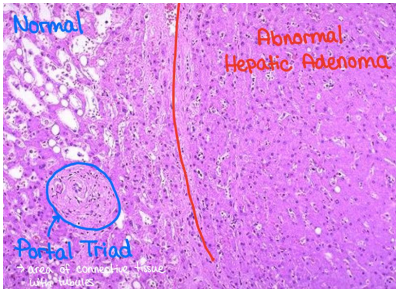
This is a higher magnification image of the same tumor. On the left is normal liver tissue with a portal tract and steatosis (white fat droplets). The abnormal tissue has a darker pink/purple color from the increase in fibrosis which compresses the sinusoids (white stripes). The adenoma cells closely resemble normal hepatocytes, but the neoplastic liver tissue is disorganized and does not contain a normal lobular architecture.
Malignant Tumors
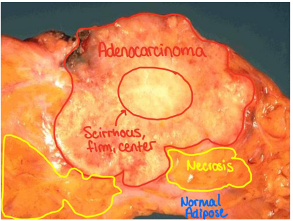
Adenocarcinoma
Breast Tissue
An irregular mass lesion invading the basement membrane (ductal carcinoma). Dense desmoplasia at the center makes it very firm (scirrhous) and white. Areas of yellowish necrosis can be seen in the portions of neoplasm invading the surrounding normal adipose tissue.
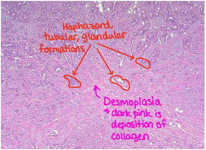
Ductal Carcinoma
Breast tissue
A central area of desmoplasia indicated by the dark pink collagen deposition. This collagen is responsible for the rigid, scirrhous nature which is palpable. The radiating outgrowth of the collagen deposition invades surrounding ductal breast tissue and is classified as malignant infiltrating ductal carcinoma.
Such an invasive carcinoma may be fixed to underlying chest wall, which also makes it immobile.
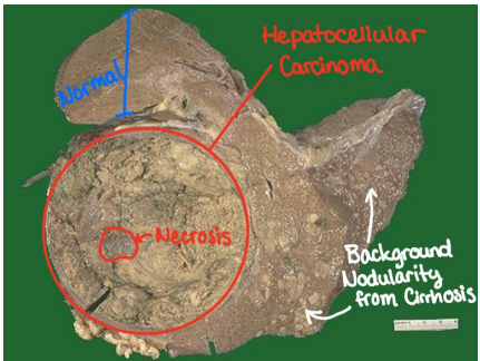
Hepatocellular Carcinoma
Cut surface of liver
The malignant hepatocellular carcinoma is the bulky neoplasm with greenish hue observed on the left invading the surrounding tissue. The greenish color most likely is due to bile production, and the grey splotches indicate necrosis.
This presentation can often be seen in viral hepatitis and chronic alcoholism.
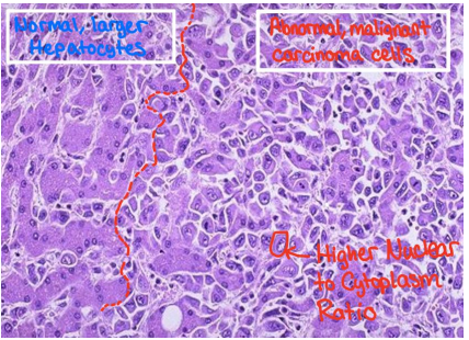
This is a higher magnification view of the same tumor. On the left is normal, large, well defined hepatocytes with small, round nuclei. On the right, is well differentiated malignant cells of this hepatocellular carcinoma. These malignant cells interdigitate with the normal hepatocytes and can be identified by their increased nuclear size causing a higher nucleus to cytoplasm ratio.
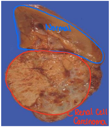
Renal Cell Carcinoma
Kidney
A fairly well circumscribed neoplasm arising in the bottom half of the cut surface of the kidney. The appearance is variegated with yellowish areas, white areas, brown areas, and hemorrhagic red areas.
Symptoms will include lower back pain, bleeding (hematuria), mass effect.
This case is heavy cigarette smoking.
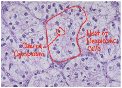
This is a higher magnification view of the same tumor. Often called “Clear Cell Carcinoma” because of the characteristic clear cytoplasm of the neoplastic cells in malignant renal cell carcinomas. The neoplastic cells abnormally shaped and arranged into nests with intervening blood vessels.
Note: In this image, the nucleus to cytoplasm ratio does not appear high because we are looking at a low-grade portion of the renal cell carcinoma
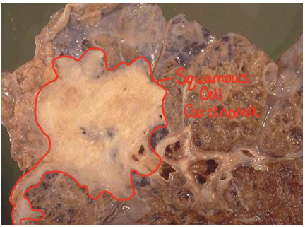
Squamous Cell Carcinoma
Lung
This type of malignant squamous cell carcinoma typically arises from squamous metaplasia. It often develops near the hiliar region and extends to the pleura. This can lead to bronchiole obstruction.
Symptoms include bleeding, productive bloody cough, bronchus obstruction often leading to secondary pneumonia,
This case was caused by smoking leading to dysplasia and then neoplasia.
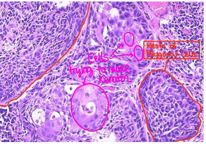
This is a higher magnification view of the same tumor. This malignant squamous cell carcinoma has nests of polygonal cells with distinct cell borders. Increased nucleus to cytoplasm ratio and hyperchromatic, dark, angular nuclei can be seen. Some cells that are trying to continue making keratin, but as cells get further from looking normal, they go from moderate to poor differentiation.
Osteosarcoma
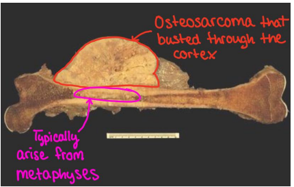
Femur
An abnormal, malignant mass that most likely arose from the metaphyseal region and then busted through the cortex of the femur. Because bone cells are not of epithelial origin, “carci” is not used. Instead, “sarco” is used because of neoplasms mesenchymal derivation.
This case was mitotic activity of growth spurts, which provides more opportunities for mutation.
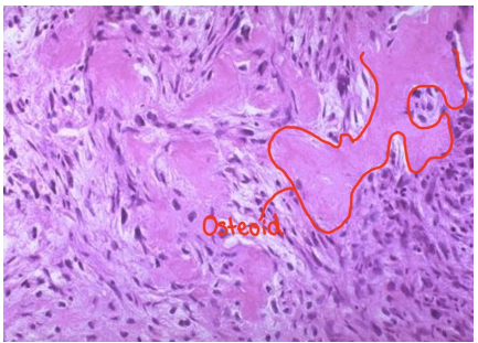
This is a higher magnification view of the same tumor. A smooth pink material called osteoid is being produced by the neoplastic spindle cells of the osteosarcoma. This osteoid production is diagnostic of osteosarcomas.
Melanoma
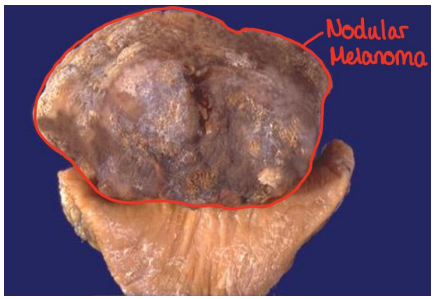
A large, dark, pigmented, nodular malignant melanoma can be seen. This is an abnormal appearance of a melanoma as they normally appear flat and are not nodular.
This is a higher magnification view of the same tumor. This image shows “giant cells” which are polygonal and have prominent nucleoli and pleiomorphic nuclei. The brown pigment visible is melanin being produced by the neoplastic melanocytes.
Lymphoma
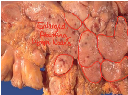
Mesenteric Lymph Nodes
This image shows a cross section of the mesentery (GI Tract) presenting with multiple, enlarged lymph nodes abutting one another. In lymphoma, lymph nodes typically appear to maintain a fleshy, tan appearance unlike the necrosis seen in metastases.
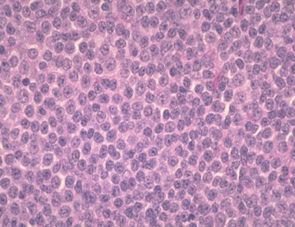
This higher magnification view reveals polygonal cells and hyperchromatic nuclei.
Metastasis
Extensive Metastases
Liver
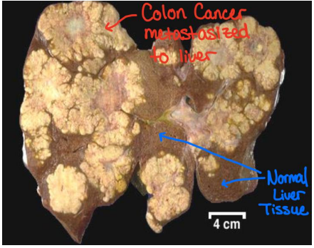
While a primary neoplasm will be a single mass, the diffuse lesions invading the normal brown colored liver tissue indicate metastases. Metastases is the best indicator of neoplastic malignancy.
Adenocarcinoma
Lymph Node
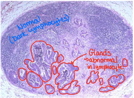
Normally characterized by a high concentration of lymphocytes with small and dark appearance. In this image, the glandular growth is an abnormal histological presentation for lymph nodes, suggesting metastasis from elsewhere since lymph nodes don’t have glands. It is common for carcinomas to metastasize to lymph nodes.
